General Information About Laryngeal Cancer
- Laryngeal cancer arises from the tissues of the larynx
- Smoking and alcohol increase the risk of laryngeal cancer
- Signs and symptoms of laryngeal cancer include a sore throat, hoarse voice, earache, and difficulty in swallowing
Laryngeal Cancer arises from the tissues of the larynx.
The larynx (voice box) lies between the base of the tongue and the trachea. The larynx contains the vocal cords, which vibrate and create sound when air passes through them. The sound echoes through the pharynx, mouth, and nose to make a person’s voice.
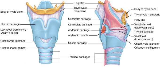
There are three main parts of the larynx:
- Supraglottis: The upper part of the larynx above the vocal cords
- Glottis: The middle part of the larynx which contains the vocal cords
- Subglottis: The lower part of the larynx between the vocal cords and the trachea (windpipe)
Smoking and alcohol excess are the primary risk factors for laryngeal cancer.
Risk factors for hypopharyngeal cancer include:
- Smoking
- Alcohol excess
Laryngeal cancer usually presents with a sore throat, hoarse voice, difficulties in swallowing and earache.
Check with your doctor if you have any of the following:
- A sore throat that does not go away.
- A lump in the neck.
- Painful or difficult swallowing.
- A change in voice (progressive hoarseness).
Examination, Diagnosis, Investigations
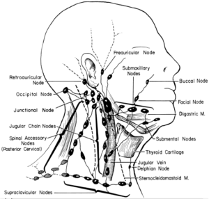
- Clinical examination and history: Your OMFS H&N surgeon will ask about your social habits (smoking and drinking) and take a thorough medical history. The neck will be felt for swollen lymph nodes.
- Fiberoptic nasoendoscopy: Your OMFS H&N surgeon will pass a thin tube-like camera through your nose, to examine the back of your throat, the base of your tongue and your voice box (larynx). This is the first examination that will allow your doctor to directly visualise if you have laryngeal cancer.
- Biopsy: This is paramount in order to diagnose cancer. Your OMFS H&N Surgeon will take a sample of the abnormal area. Given the difficulty in getting to this particular area of the throat, you will need to have an EUA (Examination Under Anaesthesia), and a direct laryngoscopy
- Fine Needle Aspiration: This is a form of biopsy, but instead of tissue sample, the OMFS H&N Surgeon samples few cells with the help of a syringe and needle. This is usually the case for lumps in the neck (lymph nodes). The cells are viewed under a microscope to find out if they are abnormal.
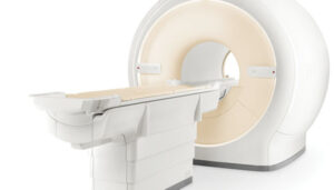
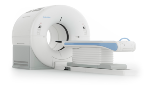
- MRI (Magnetic Resonance Imaging): A procedure that uses a magnet, radio waves, and a computer to make a series of detailed pictures of areas inside the body. An MRI of your H&N region is required to stage the cancer.
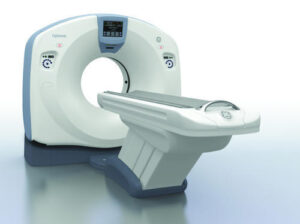
- CT scan: A computerised x-ray that makes a series of detailed pictures of areas inside the body, taken from different angles. A CT scan of your thorax (chest) is needed for staging H&N cancer, to exclude disease spread and/or second primaries in the lung. In cases of laryngeal cancer, the CT scan is important to identify whether the cancer has spread into the cartilage of your larynx (voice box) (F5).
- PET scan (positron emission tomography scan): A procedure to find malignant tumour cells in the body. A small amount of radioactive glucose (sugar) is injected into a vein. The PET scanner takes picture of where glucose is being used in the body. Malignant tumour cells show up brighter in the picture because they are more active and take up more glucose than normal cells do.
- EUA (Examination Under Anaesthesia): For this exam, you will be asleep. Your OMFS H&N surgeon will thoroughly examine the areas of your throat and take samples from abnormal looking areas. During this procedure, you will have a direct laryngoscopy, using special scopes to look at your voice box.
- Barium swallow: For that test, you will drink a liquid that contains Barium, which allows for a special X-ray to be taken, that assesses your swallow and whether there are any blockages in the course.
Factors affecting the outcome
The prognosis (chance of cure and recovery) depends on:
- The fitness of the patient
- Whether the patient is/was a smoker
- The stage of the cancer
- The location of the cancer
Stages of laryngeal Cancer
- The most important question to answer during staging is whether the cancer has spread to the cartilage of the larynx.
- There are three ways that cancer spreads in the body.
- Cancer may spread from where it began to other parts of the body.
- The following stages are used for laryngeal cancer:
- Stage 0 (Carcinoma in Situ)
- Stage I
- Stage II
- Stage III
- Stage IV
The way laryngeal cancer is staged is complex – your OMFS H&N surgeon will explain the stage and the implications once all tests are completed.
The process used to find out if laryngeal cancer has spread to other parts of the body is called staging. Staging guides decision-making and treatment.
There are three main ways that cancer spreads in the body.
Cancer can spread through tissue, the lymph system, and the blood:
- The cancer spreads from where it began by growing into nearby areas.
- Lymph system. The cancer spreads to the lymph glands (in H&N cancer it spreads in the neck lymph glands).
- The cancer spreads from where it began by getting into the blood. The cancer travels through theblood vessels to other parts of the body (distant metastasis).
When cancer spreads to another part of the body, it is called metastasis.
Treatment
- Because the larynx is the organ of breath and phonation, every effort should be made to preserve it, if possible.
- Surgery is reserved either for very early laryngeal cancers (TOLS – laser resection) or very advanced, that invade the cartilage of the larynx.
- For advanced laryngeal cancers or for cancers that do not respond to radiotherapy a procedure called laryngectomy is carried out. This removes the voice box and results in a permanent tracheostomy.
- If the laryngeal cancer has spread outside the voice box to the hypopharynx, a procedure called pahryngolaryngectomy is carried out. This removes the voice box and the part of the hypopharynx that has the cancer, and often requires reconstruction of the part of the throat that has been removed.
- Radiotherapy with or without chemo is the first line treatment for those laryngeal cancers that do not involve the cartilage of the larynx.
- TOLS and radiotherapy are equally effective for early stage laryngeal cancers, but post-treatment voice quality might be different.
- Salvage surgery is offered when laryngeal cancer doesn’t respond to radiotherapy.
- Recent advances in surgery (Transoral Laser resection – TOLS and Transoral Robotic resection – TORS) are increasingly used.
- Patients with laryngeal cancer should have their treatment planned and delivered by a team who is expert in treating head and neck cancer.
- Two types of standard treatment are used:
- Surgery(best offered from surgeons experienced in TOLS or TORS)
- Radiation therapy
- Chemotherapy (only in selected cases and as adjunct to radiotherapy)
- Treatment for laryngeal cancer is difficult, complex and may cause side effects.
- Follow-up tests may be needed.
Patients with laryngeal cancer should have their treatment planned by a team of doctors who are expert in treating head and neck cancer.
Your expert OMFS/H&N Surgeon, who will guide the team for you, will oversee your treatment. The larynx (voice box), neck and face are vital areas for eating, drinking, talking, cosmesis and psychology. A team approach is required to overcome the potential side effects of the treatment. The team usually include:
- Head and Neck Surgeon (ablation and reconstruction)
- Radiation oncologist
- Speech therapist
- Dietitian
- Pathologist
- Radiologist
- CNS
Surgery
Surgery (removing the cancer in an operation) can be considered when appropriate. Surgery might include the following:
- TOLS or TORS resection of the primary tumour: With this technique, your OMFS H&N Surgeon will use laser or a robot, to remove the primary cancer from areas that are difficult to access otherwise. This is a difficult technique that requires extensive training and skill; but it has very low morbidity and in most cases doesn’t need reconstruction of the surgical defect. It is suggested that it might lead to better swallowing compared to traditional treatment modalities. This technique is offered to patients with very small early tumours of the larynx.
- Total Laryngectomy: A procedure to remove the larynx (voice box) when the cancer has invaded the cartilage of the larynx or when the cancer hasn’t responded to the radiotherapy.
- Partial laryngectomy procedures: A variety of surgical procedures that remove parts of the larynx – getting out of fashion with the recent advances in radiotherapy, robotics, and laser surgery.
- Neck dissection: Removal of lymph nodes in the neck. This is done when cancer has or may have spread from the lip and oral cavity. This also provides access to blood vessels for microvascular reconstruction.
- Reconstruction: In cases that the laryngeal cancer has spread to the cartilage of the larynx and to the hypopharynx, a procedure called pharyngolaryngectomy is carried out. This removes the voice box and the part of the hypopharynx that has the cancer. You will have a permanent tracheostomy that will allow you to breathe safely, and, if possible, you will have a valve to facilitate your speech. You will most likely require reconstruction of your pharynx, and your OMFS Surgeon will discuss reconstruction options with you. This is a highly demanding and highly important part of laryngeal cancer treatment. The defect created by the removal of the tumour needs to be re-build to improve function and reduce the risk of potential side effects. The gold standard is the use of microvascular free tissue transfer (the surgeon takes tissue – skin, muscle, or combinations – from other parts of the patient’s’ body, preserving the blood vessels and re-perfuses the flap by anastomosing the blood vessels with donor vessels in the neck. This is called microvascular surgery and it is done under the operating microscope). Offering this kind of treatment should be the minimum you would expect from your OMFS H&N Surgeon.
Radiotherapy
Radiotherapy is a cancer treatment that uses high-energy radiation to kill cancer cells or keep them from growing. For laryngeal cancers, it can be used alone or in a combination with chemotherapy as the single required treatment, for the majority of the cases (see above for indications for surgery). Sometimes, radiotherapy is given after the surgery, depending on specific indications, and is called adjuvant therapy. Occasionally, in the event of residual tumour after surgery or if there is disease in the neck glands with extracapsular spread (ECS), radiotherapy will be given combined with chemotherapy, to increase its efficacy.
Side Effects
Treatment for mouth cancer is difficult and can result in life-changing side effects. Your OMFS H&N surgeon will follow you up and try to deal with these side effects whenever possible
- Xerostomia – dry mouth
- Difficulties in speech
- Difficulties in swallowing (may require feeding tube)
- Changes in appearance
- Weight loss
- Infection
- Lymphedema
- Chronic pain
- Osteoradionecrosis (ORN)
- Shoulder dysfunction
- Taste changes
- Scarring
- Permanent tracheostomy (stoma)
- Speaking valve